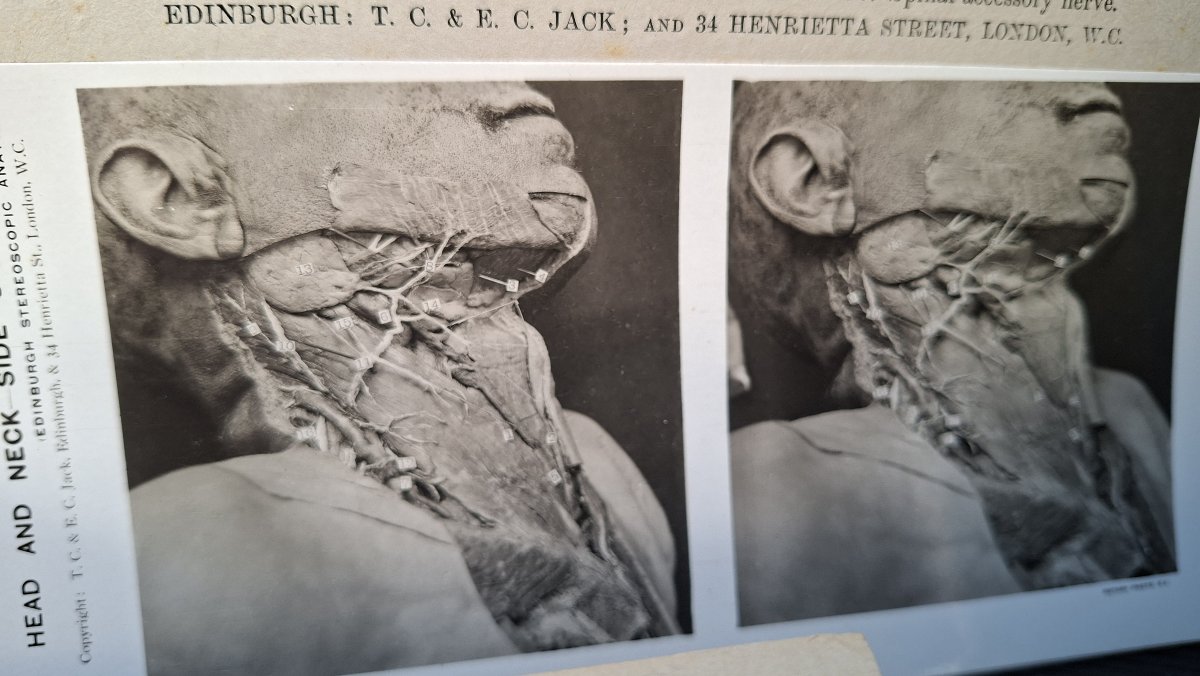
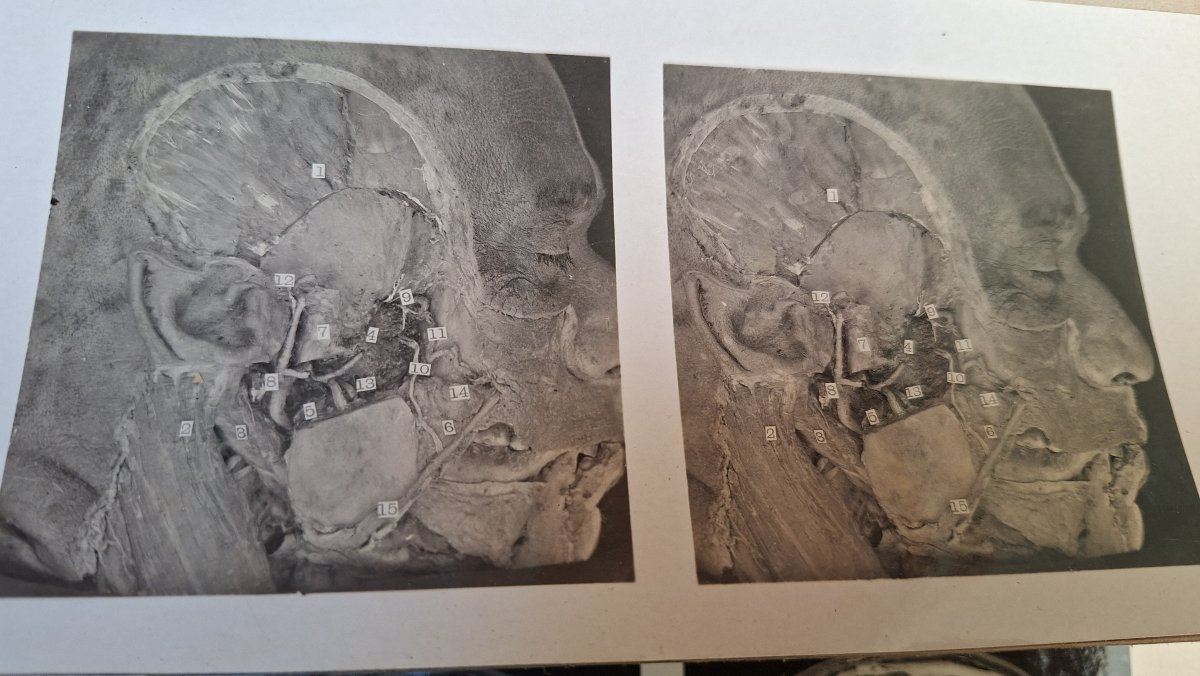
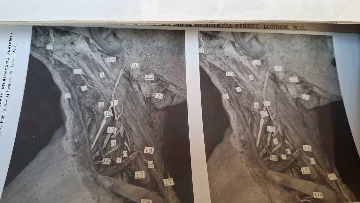
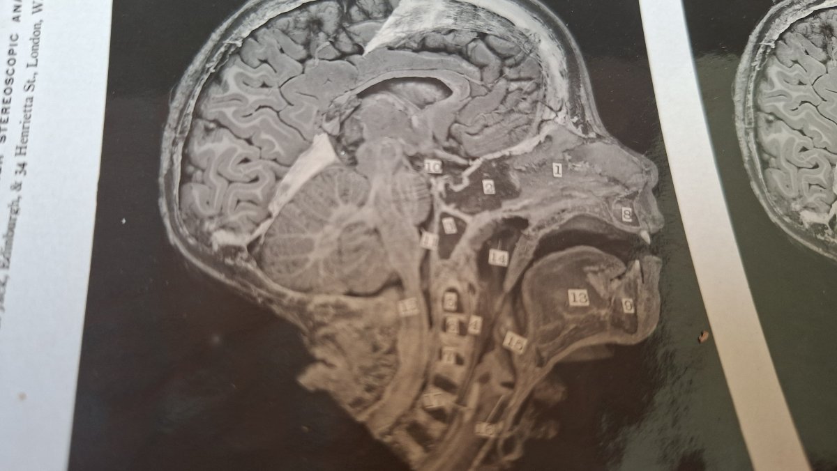
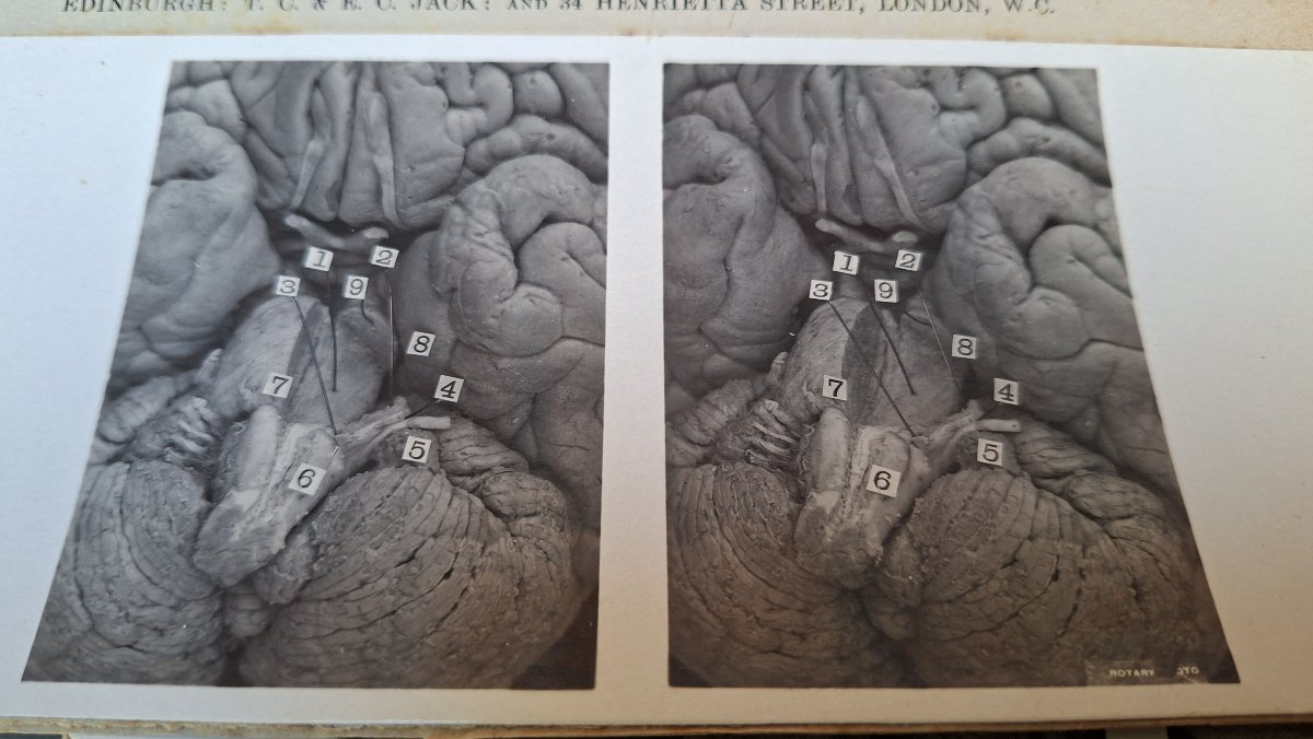
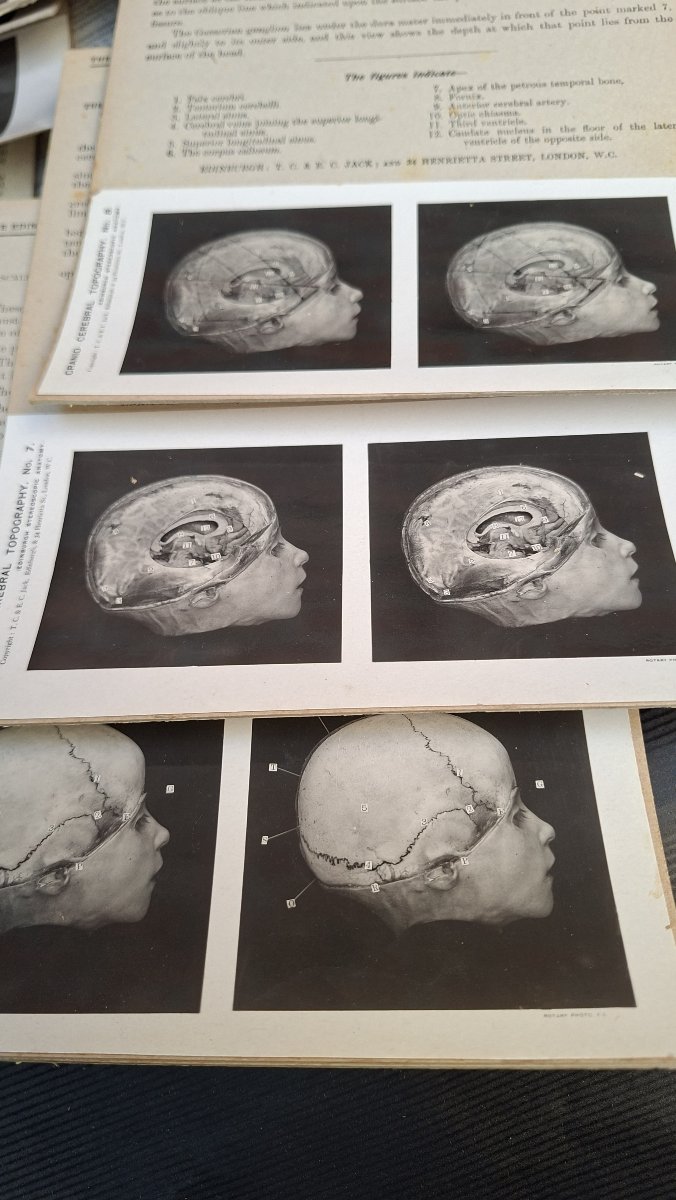
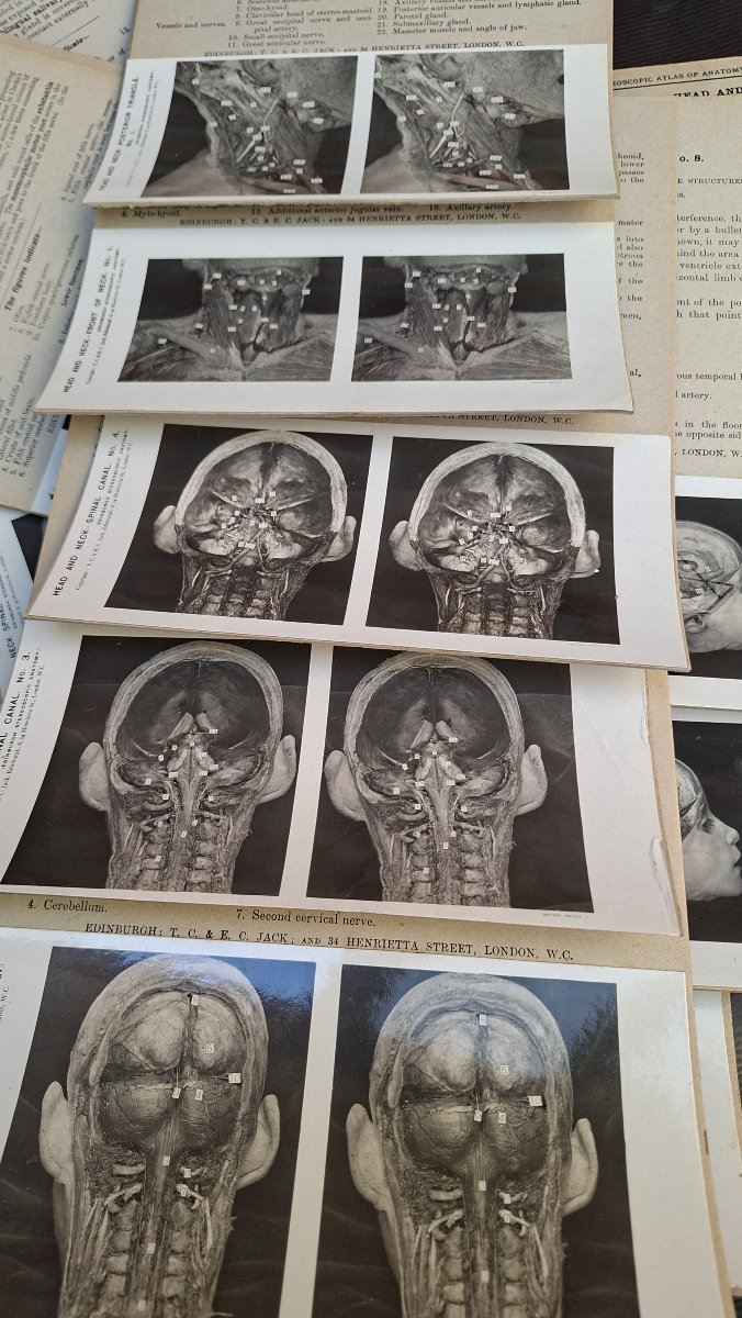
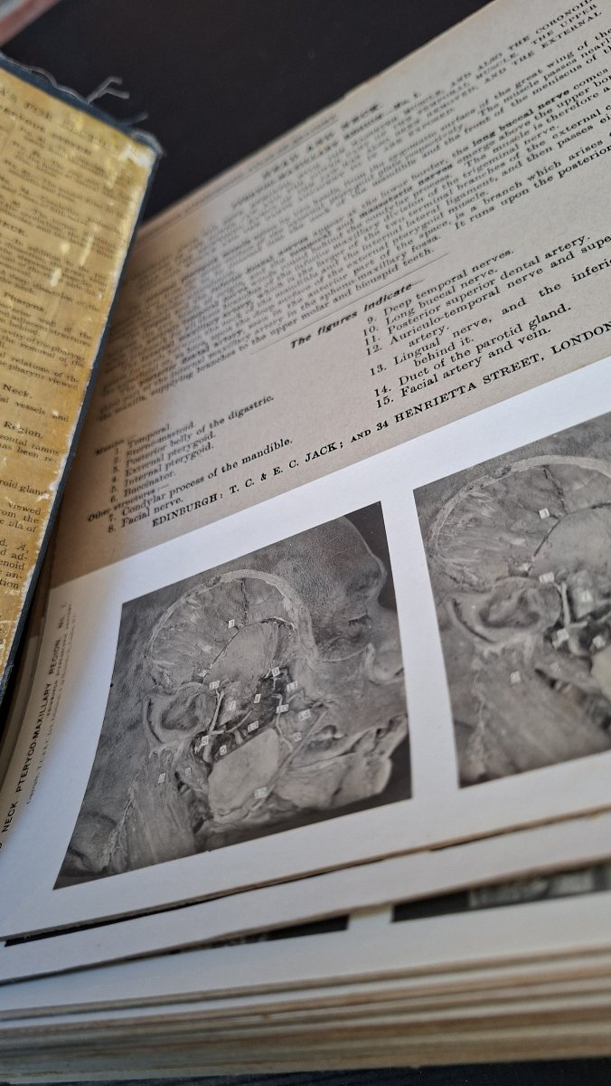
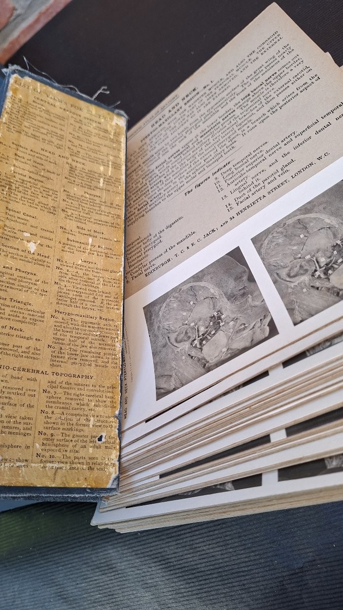
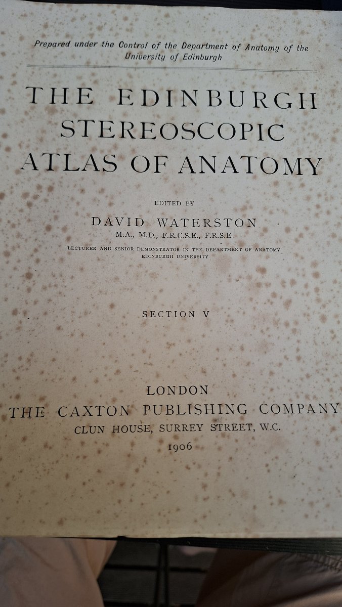


3d Atlas Of The Brain, Edinburgh, 1906, Dissection
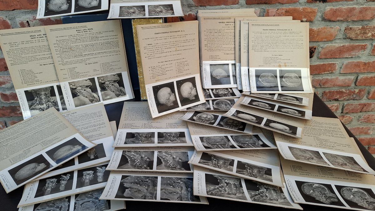
A tool for students, professionals, or knowledgeable amateurs. Immersing yourself in 3D views of brain autopsies on dissected cadavers may not be to everyone’s taste.
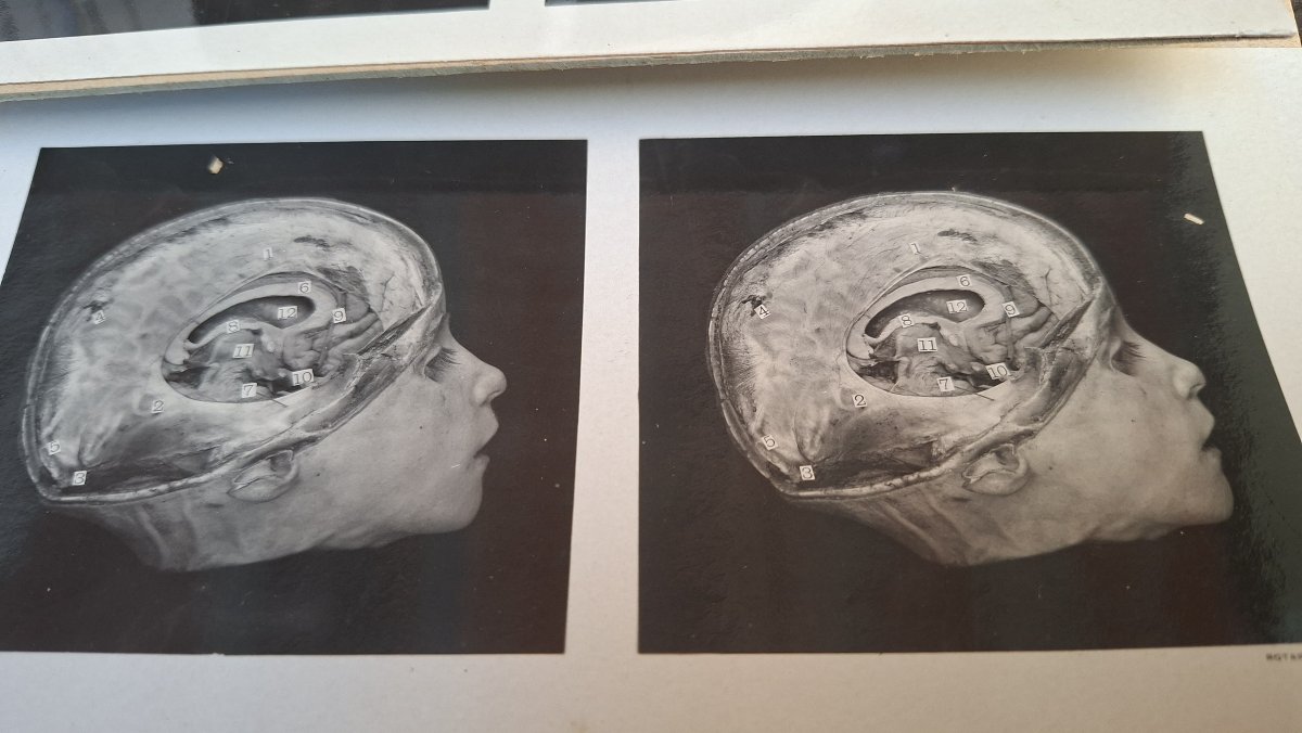
There are even images of children’s skulls.....Ah, science—it certainly has its merits.
So Volume V, the section on the brain, is complete.
The labels on the photos are written in English, which is normal in Edinburgh, even in 1906.
There are even images of children’s skulls.....Ah, science—it certainly has its merits.
So Volume V, the section on the brain, is complete.
The labels on the photos are written in English, which is normal in Edinburgh, even in 1906.
285 €
Period: 20th century
Style: Other Style
Condition: Excellent condition
Material: Other
Reference (ID): 1822695
Availability: In stock
Print